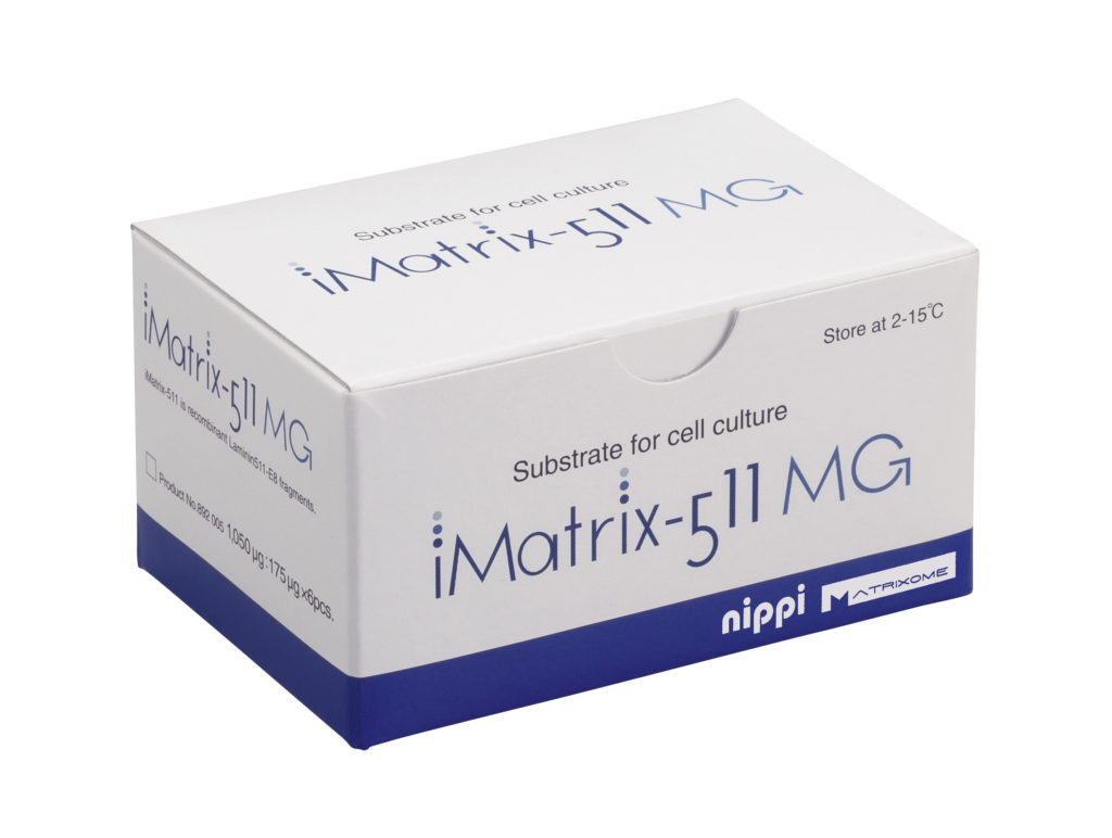
iMatrix-511MG
| Availability | In Stock |
|---|---|
| Price | Contact Us |
| Shipping | Contact Us |
| Pack Sizes | 6 x 175μg (1050μg) |
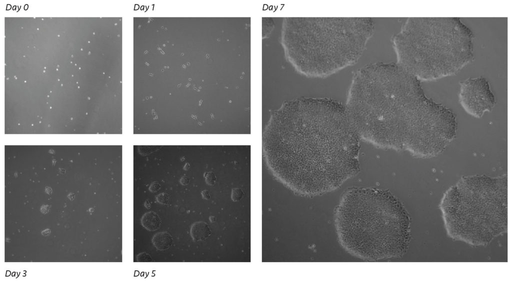
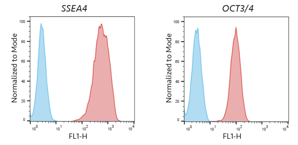
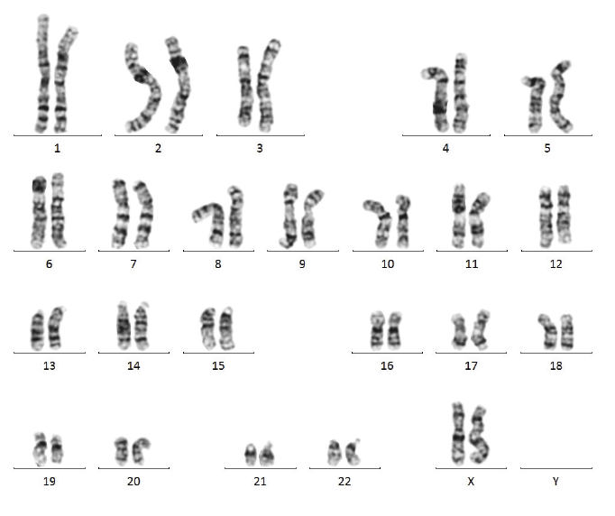
Data:
 |
 |
 *Shipments may take longer due to customs and duty clearances/inspections.
Related News:
With well over 200+ publications and citations and quickly growing, iMatrix™ products are always being used in new and novel ways leading to many breakthroughs and developments: |
 |
If you have any more questions please visit our FAQ or contact us directly using our online form here.
For more references and citations please click here.
iMatrix-511MG
iMatrix-511
iMatrix-511MG
| Citation | PMID |
|---|---|
Overview: an iPS cell stock at CiRA.
Umekage, M; Sato, Y; Takasu, N
Inflammation and Regeneration. 2019 ; 17. Doi: 10.1186/s41232-019-0106-0AbstractInduced pluripotent stem cells (iPSCs) can be produced from various somatic cells and have the ability to differentiate into various cells and tissues of the body. Regenerative medicine using iPSCs is expected to manage diseases lacking effective treatments at present. We are establishing a safe and effective iPSC stock that can be used for regenerative medicine. Our iPSC stock is recruited from healthy, consenting HLA-type homozygous donors and is made with peripheral blood-derived mononuclear cells or umbilical cord blood. We hope to minimize the influence of immune rejection by preparing HLA homozygous iPSCs. Our stock is made at the Cell Processing Center (CPC), Center for iPS Cell Research and Application (CiRA). We are preparing iPS cells that maximize matching of the Japanese population at the major HLA loci. This iPSC stock is intended to be offered not only to Japanese centers but also overseas medical institutions and companies. In August 2015, we began offering the iPSC stock for regenerative medicine and now offer 21 clones made from 5 donors. |
31497180 |
Pre-clinical study of induced pluripotent stem cell-derived dopaminergic progenitor cells for Parkinson’s disease
Doi D, Magotani H, Kikuchi T, Ikeda M, Hiramatsu S, Yoshida K, Amano N, Nomura M, Umekage M, Morizane A, Takahashi J.
Nat Commun. 2020 Jul 6;11(1):3369. Doi: 10.1038/s41467-020-17165-w.AbstractInduced pluripotent stem cell (iPSC)-derived dopaminergic (DA) neurons are an expected source for cell-based therapies for Parkinson's disease (PD). The regulatory criteria for the clinical application of these therapies, however, have not been established. Here we show the results of our pre-clinical study, in which we evaluate the safety and efficacy of dopaminergic progenitors (DAPs) derived from a clinical-grade human iPSC line. We confirm the characteristics of DAPs by in vitro analyses. We also verify that the DAP population include no residual undifferentiated iPSCs or early neural stem cells and have no genetic aberration in cancer-related genes. Furthermore, in vivo studies using immunodeficient mice reveal no tumorigenicity or toxicity of the cells. When the DAPs are transplanted into the striatum of 6-OHDA-lesioned rats, the animals show behavioral improvement. Based on these results, we started a clinical trial to treat PD patients in 2018. |
32632153 |
| Citation | PMID |
|---|---|
Characterization of dystroglycan binding in adhesion of human induced pluripotent stem cells to laminin-511 E8 fragment.
Sugawara, Y; Hamada, K; Yamada, Y; Kumai, J; Kanagawa, M; Kobayashi, K; Toda, T; Negishi, Y; Katagiri, F; Hozumi, K; Nomizu, M; Kikkawa, Y
Scientific Reports. 2019 Sep; 13037. Doi: 10.1038/s41598-019-49669-xAbstractHuman induced pluripotent stem cells (hiPSCs) grow indefinitely in culture and have the potential to regenerate various tissues. In the development of cell culture systems, a fragment of laminin-511 (LM511-E8) was found to improve the proliferation of stem cells. The adhesion of undifferentiated cells to LM511-E8 is mainly mediated through integrin α6β1. However, the involvement of non-integrin receptors remains unknown in stem cell culture using LM511-E8. Here, we show that dystroglycan (DG) is strongly expressed in hiPSCs. The fully glycosylated DG is functionally active for laminin binding, and although it has been suggested that LM511-E8 lacks DG binding sites, the fragment does weakly bind to DG. We further identified the DG binding sequence in LM511-E8, using synthetic peptides, of which, hE8A5-20 (human laminin α5 2688-2699: KTLPQLLAKLSI) derived from the laminin coiled-coil domain, exhibited DG binding affinity and cell adhesion activity. Deletion and mutation studies show that LLAKLSI is the active core sequence of hE8A5-20, and that, K2696 is a critical amino acid for DG binding. We further demonstrated that hiPSCs adhere to hE8A5-20-conjugated chitosan matrices. The amino acid sequence of DG binding peptides would be useful to design substrata for culture system of undifferentiated and differentiated stem cells. |
31506597 |
Numerical operations in living cells by programmable RNA devices.
Endo, K; Hayashi, K; Saito, H
Science Advances. 2019 Aug; eaax0835. Doi: 10.1126/sciadv.aax0835AbstractIntegrated bioengineering systems can make executable decisions according to the cell state. To sense the state, multiple biomarkers are detected and processed via logic gates with synthetic biological devices. However, numerical operations have not been achieved. Here, we show a design principle for messenger RNA (mRNA) devices that recapitulates intracellular information by multivariate calculations in single living cells. On the basis of this principle and the collected profiles of multiple microRNA activities, we demonstrate that rationally programmed mRNA sets classify living human cells and track their change during differentiation. Our mRNA devices automatically perform multivariate calculation and function as a decision-maker in response to dynamic intracellular changes in living cells. |
31457099 |
Using the Inducible Caspase-9 Suicide-Safeguard System with iPSC and Bioluminescent Tracking.
Villanueva, J; Nishimura, T; Nakauchi, H
Methods in Molecular Biology. 2019 ; 259-264. Doi: 10.1007/978-1-4939-9728-2_20AbstractFor scientists working within the field of induced pluripotent stem cells (iPSCs), this protocol will provide a thorough walk-through on how to conduct in vitro and in vivo experiments that validate the function of a particular safeguard system technology. In short, we provide instructions on how to generate inducible Caspase-9 (iC9) safeguard system with human iPSCs that act as normal or abnormal models of the cells for therapeutics to be tried after differentiation. These iC9-iPSCs should be modified prior to beginning this protocol by constitutively expressing luciferase, an enzyme capable of generating bioluminescent signals through the oxidation of the substrate luciferin. Monitoring the bioluminescent signal over time provides the information on whether a safeguard system is working or not. |
31396943 |
In Vitro Differentiation of T Cell: From CAR-Modified T-iPSC.
Ueda, T; Kaneko, S
Methods in Molecular Biology. 2019 ; 85-91. Doi: 10.1007/978-1-4939-9728-2_10AbstractT cells engineered to express chimeric antigen receptor (CAR) against the B cell antigen CD19 are achieving remarkable clinical effects on hematological malignancies. Allogeneic transplantation approach is promising for broaden application of CART therapy. iPSCs are one of the ideal cell sources for this approach. CAR-engineered iPSCs are demonstrated to give rise to CAR-engineered T cell and exert their effector function. In this section, we describe the method to generate CAR-engineered iPSCs and differentiate them into T cells. |
31396933 |
Differentiating CD8αβ T Cells from TCR-Transduced iPSCs for Cancer Immunotherapy.
Minagawa, A; Kaneko, S
Methods in Molecular Biology. 2019 ; 81-84. Doi: 10.1007/978-1-4939-9728-2_9AbstractThe use of induced pluripotent stem cells (iPSCs) as a cell source for producing cytotoxic T lymphocytes (CTLs) is expected to have advantages in the antigen specificity, rejuvenation profile, and reproducible number of CTLs. We have developed the way to differentiate CD8αβ T cells from TCR-transduced iPSCs (TCR-iPSCs). These T cells express monoclonal expression of the transduced TCR. Generating CD8αβ CTLs from TCR-iPSC could contribute to safe and effective allogeneic regenerative T cell immunotherapies. |
31396932 |
Human iPSC Generation from Antigen-Specific T Cells.
Nishimura, T; Murmann, Y; Nakauchi, H
Methods in Molecular Biology. 2019 ; 53-57. Doi: 10.1007/978-1-4939-9728-2_5AbstractThe discovery and development of induced pluripotent stem cells (iPSCs) opened a novel venue for disease modeling, drug discovery, and personalized medicine. Additionally, iPSCs have been utilized for a wide variety of research and clinical applications without immunological and ethical concerns that encounter embryonic stem cells. Adoptive T cell immunotherapy is a form of cellular immunotherapy that involves transfusion of functional T cells. However, this approach requires T cell expansion and the process causes T cell exhaustion. As a result, highly expanded T cells have not proven to be particularly effective for treatments. This exhaustion issue could be overcome due to rejuvenation of T cells by reprogramming to pluripotency and redifferentiation to T cells. This is a potential therapeutic strategy for combating various types of cancer. |
31396928 |
Establishment of human trophoblast stem cells from human induced pluripotent stem cell-derived cystic cells under micromesh culture.
Li, Z; Kurosawa, O; Iwata, H
Stem Cell Research & Therapy. 2019 Aug; 245. Doi: 10.1186/s13287-019-1339-1AbstractTrophoblasts as a specific cell lineage are crucial for the correct function of the placenta. Human trophoblast stem cells (hTSCs) are a proliferative population that can differentiate into syncytiotrophoblasts and extravillous cytotrophoblasts. Many studies have reported that chemical supplements induce the differentiation of trophoblasts from human induced pluripotent stem cells (hiPSCs). However, there have been no Reports of the establishment of proliferative hTSCs from hiPSCs. Our previous report showed that culturing hiPSCs on micromesh as a bioscaffold induced cystic cells with trophoblast properties. Here, we aimed to establish hTSCs from hiPSCs.We used the micromesh culture technique to induce hiPSC differentiation into trophoblast cysts. We then reseeded and purified cystic cells.The cells derived from the reseeded cysts were highly proliferative. Low expression levels of pluripotency genes and high expression levels of TSC-specific genes were detected in proliferative cells. The cells could be passaged, and further directional differentiation into syncytiotrophoblast- and extravillous cytotrophoblast-like cells was confirmed by marker expression and hormone secretion.We established hiPSC-derived hTSCs, which may be applicable for studying the functions of trophoblasts and the placenta. Our experimental system may provide useful tools for understanding the pathogenesis of infertility owing to trophoblast defects in the future. |
31391109 |
Activin Is Superior to BMP7 for Efficient Maintenance of Human iPSC-Derived Nephron Progenitors.
Tanigawa, S; Naganuma, H; Kaku, Y; Era, T; Sakuma, T; Yamamoto, T; Taguchi, A; Nishinakamura, R
Stem Cell Reports. 2019 Aug; 322-337. Doi: 10.1016/j.stemcr.2019.07.003AbstractKidney formation is regulated by the balance between maintenance and differentiation of nephron progenitor cells (NPCs). Now that directed differentiation of NPCs from human induced pluripotent stem cells (iPSCs) can be achieved, maintenance and propagation of NPCs in vitro should be beneficial for regenerative medicine. Although WNT and FGF signals were previously shown to be essential for NPC propagation, the requirement for BMP/TGFβ signaling remains controversial. Here we reveal that activin has superior effects to BMP7 on maintenance efficiency of human iPSC-derived NPCs. Activin expanded ITGA8+/PDGFRA-/SIX2-GFP+ NPCs by 5-fold per week at 80%-90% efficiency, and the propagated cells possessed robust capacity for nephron formation both in vitro and in vivo. The expanded cells also maintained their nephron-forming potential after freezing. Furthermore, the protocol was applicable to multiple non-GFP-tagged iPSC lines. Thus, our activin-based protocol will be applicable to a variety of research fields including disease modeling and drug screening. |
31378669 |
Directed differentiation of human induced pluripotent stem cells into mature stratified bladder urothelium.
Suzuki, K; Koyanagi-Aoi, M; Uehara, K; Hinata, N; Fujisawa, M; Aoi, T
Scientific Reports. 2019 Jul; 10506. Doi: 10.1038/s41598-019-46848-8AbstractFor augmentation or reconstruction of urinary bladder after cystectomy, bladder urothelium derived from human induced pluripotent stem cells (hiPSCs) has recently received focus. However, previous studies have only shown the emergence of cells expressing some urothelial markers among derivatives of hiPSCs, and no report has demonstrated the stratified structure, which is a particularly important attribute of the barrier function of mature bladder urothelium. In present study, we developed a method for the directed differentiation of hiPSCs into mature stratified bladder urothelium. The caudal hindgut, from which the bladder urothelium develops, was predominantly induced via the high-dose administration of CHIR99021 during definitive endoderm induction, and this treatment subsequently increased the expressions of uroplakins. Terminal differentiation, characterized by the increased expression of uroplakins, CK13, and CK20, was induced with the combination of Troglitazone + PD153035. FGF10 enhanced the expression of uroplakins and the stratification of the epithelium, and the transwell culture system further enhanced such stratification. Furthermore, the barrier function of our urothelium was demonstrated by a permeability assay using FITC-dextran. According to an immunohistological analysis, the stratified uroplakin II-positive epithelium was observed in the transwells. This method might be useful in the field of regenerative medicine of the bladder. |
31324820 |
Geometrical Patterning and Constituent Cell Heterogeneity Facilitate Electrical Conduction Disturbances in a Human Induced Pluripotent Stem Cell-Based Platform: An In vitro Disease Model of Atrial Arrhythmias.
Nakanishi, H; Lee, JK; Miwa, K; Masuyama, K; Yasutake, H; Li, J; Tomoyama, S; Honda, Y; Deguchi, J; Tsujimoto, S; Hidaka, K; Miyagawa, S; Sawa, Y; Komuro, I; Sakata, Y
Frontiers in Physiology. 2019 ; 818. Doi: 10.3389/fphys.2019.00818AbstractEctopic foci from pulmonary veins (PVs) comprise the main trigger associated with the initiation of atrial fibrillation (AF). An abrupt anatomical narrow-to-wide transition, modeled as in vitro geometrical patterning with similar configuration in the present study, is located at the junction of PVs and the left atrium (LA). Complex cellular composition, i.e., constituent cell heterogeneity, is also observed in PVs and the PVs-LA junction. High frequency triggers accompanied with anatomical irregularity and constituent cell heterogeneity provoke impaired conduction, a prerequisite for AF genesis. However, few experiments investigating the effects of these factors on electrophysiological properties using human-based cardiomyocytes (CMs) with atrial properties have been reported. The aim of the current study was to estimate whether geometrical patterning and constituent cell heterogeneity under high frequency stimuli undergo conduction disturbance utilizing an in vitro two-dimensional (2D) monolayer preparation consisting of atrial-like CMs derived from human induced pluripotent stem cells (hiPSCs) and atrial fibroblasts (Fbs). We induced hiPSCs into atrial-like CMs using a directed cardiac differentiation protocol with the addition of all-trans retinoic acid (ATRA). The atrial-like hiPSC-derived CMs (hiPSC-CMs) and atrial Fbs were transferred in defined ratios (CMs/Fbs: 100%/0% or 70%/30%) on manually fabricated plates with or without geometrical patterning imitating the PVs-LA junction. High frequency field stimulation emulating repetitive ectopic foci originated in PVs were delivered, and the electrical propagation was assessed by optical mapping. We generated high purity CMs with or without the ATRA application. ATRA-treated hiPSC-CMs exhibited significantly higher atrial-specific properties by immunofluorescence staining, gene expression patterns, and optical action potential parameters than those of ATRA-untreated hiPSC-CMs. Electrical stimuli at a higher frequency preferentially induced impaired electrical conduction on atrial-like hiPSC-CMs monolayer preparations with an abrupt geometrical transition than on those with uniform geometry. Additionally, the application of human atrial Fbs to the geometrically patterned atrial-like hiPSC-CMs tended to further deteriorate the integrity of electrical conduction compared with those using the atrial-like hiPSC-CM alone preparations. Thus, geometrical narrow-to-wide patterning under high frequency stimuli preferentially jeopardized electrical conduction within in vitro atrial-like hiPSC-CM monolayers. Constituent cell heterogeneity represented by atrial Fbs also contributed to the further deterioration of conduction stability. |
31316396 |
The First Generation of iPSC Line from a Korean Alzheimer's Disease Patient Carrying APP-V715M Mutation Exhibits a Distinct Mitochondrial Dysfunction.
Li, L; Roh, JH; Kim, HJ; Park, HJ; Kim, M; Koh, W; Heo, H; Chang, JW; Nakanishi, M; Yoon, T; Na, DL; Song, J
Experimental Neurobiology. 2019 Jun; 329-336. Doi: 10.5607/en.2019.28.3.329AbstractAlzheimer's Disease (AD) is a progressive neurodegenerative disease, which is pathologically defined by the accumulation of amyloid plaques and hyper-phosphorylated tau aggregates in the brain. Mitochondrial dysfunction is also a prominent feature in AD, and the extracellular Aβ and phosphorylated tau result in the impaired mitochondrial dynamics. In this study, we generated an induced pluripotent stem cell (iPSC) line from an AD patient with amyloid precursor protein (APP) mutation (Val715Met; APP-V715M) for the first time. We demonstrated that both extracellular and intracellular levels of Aβ were dramatically increased in the APP-V715M iPSC-derived neurons. Furthermore, the APP-V715M iPSC-derived neurons exhibited high expression levels of phosphorylated tau (AT8), which was also detected in the soma and neurites by immunocytochemistry. We next investigated mitochondrial dynamics in the iPSC-derived neurons using Mito-tracker, which showed a significant decrease of anterograde and retrograde velocity in the APP-V715M iPSC-derived neurons. We also found that as the Aβ and tau pathology accumulates, fusion-related protein Mfn1 was decreased, whereas fission-related protein DRP1 was increased in the APP-V715M iPSC-derived neurons, compared with the control group. Taken together, we established the first iPSC line derived from an AD patient carrying APP-V715M mutation and showed that this iPSC-derived neurons exhibited typical AD pathological features, including a distinct mitochondrial dysfunction. |
31308793 |
Long-term eradication of extranodal NK/T cell lymphoma, nasal type, by induced pluripotent stem cell-derived Epstein-Barr virus-specific rejuvenated T cells in vivo.
Ando, M; Ando, J; Yamazaki, S; Ishii, M; Sakiyama, Y; Harada, S; Honda, T; Yamaguchi, T; Nojima, M; Ohshima, K; Nakauchi, H; Komatsu, N
Haematologica. 2019 Jul; . Doi: 10.3324/haematol.2019.223511AbstractFunctionally rejuvenated induced pluripotent stem cell-derived antigen-specific cytotoxic T lymphocytes are expected to be potent immunotherapy for tumors. When L-asparaginase-containing standard chemotherapy fails in extranodal NK/T cell lymphoma, nasal type, no effective salvage therapy exists. The clinical course then is miserable. We demonstrate prolonged and robust eradication of extranodal NK/T cell lymphoma, nasal type, in vivo by Epstein-Barr virus-specific induced pluripotent stem cell-derived antigen-specific cytotoxic T lymphocytes, with induced pluripotent stem cell-derived antigen-specific cytotoxic T lymphocytes persisting as central memory T cells in mouse spleen for at least 6 months. The anti-tumor response is so strong that any concomitant effect of PD-1 blockade is unclear. These results suggest that long-term persistent Epstein-Barr virus-specific induced pluripotent stem cell-derived antigen-specific cytotoxic T lymphocytes contribute to continuous anti-tumor effect and offer an effective salvage therapy for relapsed and refractory extranodal NK/T cell lymphoma, nasal type. |
31296577 |
Dual usage of a stage-specific fluorescent reporter system based on a helper-dependent adenoviral vector to visualize osteogenic differentiation.
Sone, T; Shin, M; Ouchi, T; Sasanuma, H; Miyamoto, A; Ohte, S; Tsukamoto, S; Nakanishi, M; Okano, H; Katagiri, T; Mitani, K
Scientific Reports. 2019 Jul; 9705. Doi: 10.1038/s41598-019-46105-yAbstractWe developed a reporter system that can be used in a dual manner in visualizing mature osteoblast formation. The system is based on a helper-dependent adenoviral vector (HDAdV), in which a fluorescent protein, Venus, is expressed under the control of the 19-kb human osteocalcin (OC) genomic locus. By infecting human and murine primary osteoblast (POB) cultures with this reporter vector, the cells forming bone-like nodules were specifically visualized by the reporter. In addition, the same vector was utilized to efficiently knock-in the reporter into the endogenous OC gene of human induced pluripotent stem cells (iPSCs), by homologous recombination. Neural crest-like cells (NCLCs) derived from the knock-in reporter iPSCs were differentiated into osteoblasts forming bone-like nodules and could be visualized by the expression of the fluorescent reporter. Living mature osteoblasts were then isolated from the murine mixed POB culture by fluorescence-activated cell sorting (FACS), and their mRNA expression profile was analyzed. Our study presents unique utility of reporter HDAdVs in stem cell Biology and related applications. |
31273280 |
Generation of three induced pluripotent stem cell lines from postmortem tissue derived following sudden death of a young patient with STXBP1 mutation.
Yamamoto, T; Otsu, M; Okumura, T; Horie, Y; Ueno, Y; Taniguchi, H; Ohtaka, M; Nakanishi, M; Abe, Y; Murase, T; Umehara, T; Ikematsu, K
Stem Cell Research. 2019 Aug; 101485. Doi: 10.1016/j.scr.2019.101485AbstractWe established three iPSC lines from postmortem-cultured fibroblasts derived following the sudden unexpected death of an 8-year-old girl with Lennox-Gastaut syndrome, who turned out to have the R551H-mutant STXBP1 gene. These iPSC clones showed pluripotent characteristics while retaining the genotype and demonstrated trilineage differentiation capability, indicating their utility in disease-modeling studies, i.e., STXBP1-encephalopathy. This is the first report on the establishment of iPSCs from a sudden death child, suggesting the possible use of postmortem-iPSC technologies as an epoch-making approach for precise identification of the cause of sudden death. |
31255830 |
Intracellular Zn2+ transients modulate global gene expression in dissociated rat hippocampal neurons.
Sanford, L; Carpenter, MC; Palmer, AE
Scientific Reports. 2019 Jun; 9411. Doi: 10.1038/s41598-019-45844-2AbstractZinc (Zn2+) is an integral component of many proteins and has been shown to act in a regulatory capacity in different mammalian systems, including as a neurotransmitter in neurons throughout the brain. While Zn2+ plays an important role in modulating neuronal potentiation and synaptic plasticity, little is known about the signaling mechanisms of this regulation. In dissociated rat hippocampal neuron cultures, we used fluorescent Zn2+ sensors to rigorously define resting Zn2+ levels and stimulation-dependent intracellular Zn2+ dynamics, and we performed RNA-Seq to characterize Zn2+-dependent transcriptional effects upon stimulation. We found that relatively small changes in cytosolic Zn2+ during stimulation altered expression levels of 931 genes, and these Zn2+ dynamics induced transcription of many genes implicated in neurite expansion and synaptic growth. Additionally, while we were unable to verify the presence of synaptic Zn2+ in these cultures, we did detect the synaptic vesicle Zn2+ transporter ZnT3 and found it to be substantially upregulated by cytosolic Zn2+ increases. These results provide the first global sequencing-based examination of Zn2+-dependent changes in transcription and identify genes that may mediate Zn2+-dependent processes and functions. |
31253848 |
Phenotypic Drug Screening for Dysferlinopathy Using Patient-Derived Induced Pluripotent Stem Cells.
Kokubu, Y; Nagino, T; Sasa, K; Oikawa, T; Miyake, K; Kume, A; Fukuda, M; Fuse, H; Tozawa, R; Sakurai, H
Stem Cells Translational Medicine. 2019 Jun; . Doi: 10.1002/sctm.18-0280AbstractDysferlinopathy is a progressive muscle disorder that includes limb-girdle muscular dystrophy type 2B and Miyoshi myopathy (MM). It is caused by mutations in the dysferlin (DYSF) gene, whose function is to reseal the muscular membrane. Treatment with proteasome inhibitor MG-132 has been shown to increase misfolded dysferlin in fibroblasts, allowing them to recover their membrane resealing function. Here, we developed a screening system based on myocytes from MM patient-derived induced pluripotent stem cells. According to the screening, nocodazole was found to effectively increase the level of dysferlin in cells, which, in turn, enhanced membrane resealing following injury by laser irradiation. Moreover, the increase was due to microtubule disorganization and involved autophagy rather than the proteasome degradation pathway. These findings suggest that increasing the amount of misfolded dysferlin using small molecules could represent an effective future clinical treatment for dysferlinopathy. Stem Cells Translational Medicine2019. |
31250983 |
Robust and highly efficient hiPSC generation from patient non-mobilized peripheral blood-derived CD34+ cells using the auto-erasable Sendai virus vector.
Okumura, T; Horie, Y; Lai, CY; Lin, HT; Shoda, H; Natsumoto, B; Fujio, K; Kumaki, E; Okano, T; Ono, S; Tanita, K; Morio, T; Kanegane, H; Hasegawa, H; Mizoguchi, F; Kawahata, K; Kohsaka, H; Moritake, H; Nunoi, H; Waki, H; Tamaru, SI; Sasako, T; Yamauchi, T; Kadowaki, T; Tanaka, H; Kitanaka, S; Nishimura, K; Ohtaka, M; Nakanishi, M; Otsu, M
Stem Cell Research & Therapy. 2019 Jun; 185. Doi: 10.1186/s13287-019-1273-2AbstractDisease modeling with patient-derived induced pluripotent stem cells (iPSCs) is a powerful tool for elucidating the mechanisms underlying disease pathogenesis and developing safe and effective treatments. Patient peripheral blood (PB) cells are used for iPSC generation in many cases since they can be collected with minimum invasiveness. To derive iPSCs that lack immunoreceptor gene rearrangements, hematopoietic stem and progenitor cells (HSPCs) are often targeted as the reprogramming source. However, the current protocols generally require HSPC mobilization and/or ex vivo expansion owing to their sparsity at the steady state and low reprogramming efficiencies, making the overall procedure costly, laborious, and time-consuming.We have established a highly efficient method for generating iPSCs from non-mobilized PB-derived CD34+ HSPCs. The source PB mononuclear cells were obtained from 1 healthy donor and 15 patients and were kept frozen until the scheduled iPSC generation. CD34+ HSPC enrichment was done using immunomagnetic beads, with no ex vivo expansion culture. To reprogram the CD34+-rich cells to pluripotency, the Sendai virus vector SeVdp-302L was used to transfer four transcription factors: KLF4, OCT4, SOX2, and c-MYC. In this iPSC generation series, the reprogramming efficiencies, success rates of iPSC line establishment, and progression time were recorded. After generating the iPSC frozen stocks, the cell recovery and their residual transgenes, karyotypes, T cell receptor gene rearrangement, pluripotency markers, and differentiation capability were examined.We succeeded in establishing 223 iPSC lines with high reprogramming efficiencies from 15 patients with 8 different disease types. Our method allowed the rapid appearance of primary colonies (~ 8 days), all of which were expandable under feeder-free conditions, enabling robust establishment steps with less workload. After thawing, the established iPSC lines were verified to be pluripotency marker-positive and of non-T cell origin. A majority of the iPSC lines were confirmed to be transgene-free, with normal karyotypes. Their trilineage differentiation capability was also verified in a defined in vitro assay.This robust and highly efficient method enables the rapid and cost-effective establishment of transgene-free iPSC lines from a small volume of PB, thus facilitating the biobanking of patient-derived iPSCs and their use for the modeling of various diseases. |
31234949 |
Feasibility of a cryopreservation of cultured human corneal endothelial cells.
Okumura, N; Kagami, T; Watanabe, K; Kadoya, S; Sato, M; Koizumi, N
PloS One. 2019 ; e0218431. Doi: 10.1371/journal.pone.0218431AbstractTransparency of the cornea is essential for vision and is maintained by the corneal endothelium. Consequently, corneal endothelial decompensation arising from irreversible damage to the corneal endothelium causes severe vision impairment. Until recently, transplantation of donor corneas was the only therapeutic choice for treatment of endothelial decompensation. In 2013, we initiated clinical research into cell-based therapy involving injection of a suspension of cultured human corneal endothelial cells (HCECs), in combination with Rho kinase inhibitor, into the anterior chamber. The aim of the present study was to establish a protocol for cryopreservation of HCECs to allow large-scale commercial manufacturing of these cells. This study focused on the effects of various cryopreservation reagents on HCEC viability. Screening of several commercially available cryopreservation reagents identified Bambanker hRM as an effective agent that maintained a cell viability of 89.4% after 14 days of cryopreservation, equivalent to the cell viability of 89.2% for non-cryopreserved control cells. The use of Bambanker hRM and HCECs at a similar grade to that used clinically for cell based therapy (passage 3-5 and a cell density higher than 2000 cells/mm2) gave a similar cell density for cryopreserved HCECs to that of non-preserved control HCECs after 28 days of cultivation (2099 cells/mm2 and 2111 cells/mm2, respectively). HCECs preserved using Bambanker hRM grew in a similar fashion to non-preserved control HCECs and formed a monolayer sheet-like structure. Cryopreservation of HCECs has multiple advantages including the ability to accumulate stocks of master cells, to transport HCEC stocks, and to manufacture HCECs on demand for use in cell-based treatment of endothelial decompensation. |
31226131 |
Establishment of induced pluripotent stem cells from common marmoset fibroblasts by RNA-based reprogramming.
Nakajima, M; Yoshimatsu, S; Sato, T; Nakamura, M; Okahara, J; Sasaki, E; Shiozawa, S; Okano, H
Biochemical and Biophysical Research Communications. 2019 Aug; 593-599. Doi: 10.1016/j.bbrc.2019.05.175AbstractThe common marmoset (marmoset; Callithrix jacchus) shows anatomical and physiological features that are in common with humans. Establishing induced pluripotent stem cells (iPSCs) from marmosets holds promise for enhancing the utility of the animal model for biomedical and preclinical studies. However, in spite of the presence of some previous Reports on marmoset iPSCs, the reprogramming technology in marmosets is still under development. In particular, the efficacy of RNA-based reprogramming has not been thoroughly investigated. In this study, we attempted RNA-based reprogramming for deriving iPSCs from marmoset fibroblasts. Although we failed to derive iPSC colonies from marmoset fibroblasts by using a conventional RNA-based reprogramming method previously validated in human fibroblasts, we succeeded in deriving colony-forming cells with a modified induction medium supplemented with a novel set of small molecules. Importantly, following one-week culture of the colony-forming cells in conventional embryonic stem cell (ESC) medium, we obtained iPSCs which express endogenous pluripotent markers and show a differentiation potential into all three germ layers. Taken together, our results indicate that RNA-based reprogramming, which is valuable for deriving transgene-free iPSCs, is applicable to marmosets. |
31178141 |
F5-Peptide and mTORC1/rpS6 Effectively Enhance BTB Transport Function in the Testis-Lesson From the Adjudin Model.
Mao, B; Li, L; Yan, M; Wong, CKC; Silvestrini, B; Li, C; Ge, R; Lian, Q; Cheng, CY
Endocrinology. 2019 Aug; 1832-1853. Doi: 10.1210/en.2019-00308AbstractDuring spermatogenesis, the blood-testis barrier (BTB) undergoes cyclic remodeling that is crucial to support the transport of preleptotene spermatocytes across the immunological barrier at stage VIII to IX of the epithelial cycle. Studies have shown that this timely remodeling of the BTB is supported by several endogenously produced barrier modifiers across the seminiferous epithelium, which include the F5-peptide and the ribosomal protein S6 [rpS6; a downstream signaling molecule of the mammalian target of rapamycin complex 1 (mTORC1)] signaling protein. Herein, F5-peptide and a quadruple phosphomimetic (and constitutively active) mutant of rpS6 [i.e., phosphorylated (p-)rpS6-MT] that are capable of inducing reversible immunological barrier remodeling, by making the barrier "leaky" transiently, were used for their overexpression in the testis to induce BTB opening. We sought to examine whether this facilitated the crossing of the nonhormonal male contraceptive adjudin at the BTB when administered by oral gavage, thereby effectively improving its BTB transport to induce germ cell adhesion and aspermatogenesis. Indeed, it was shown that combined overexpression of F5-peptide and p-rpS6-MT and a low dose of adjudin, which by itself had no noticeable effects on spermatogenesis, was capable of perturbing the organization of actin- and microtubule (MT)-based cytoskeletons through changes in the spatial expression of actin- and MT-binding/regulatory proteins to the corresponding cytoskeleton. These findings thus illustrate the possibility of delivering drugs to any target organ behind a blood-tissue barrier by modifying the tight junction permeability barrier using endogenously produced barrier modifiers based on findings from this adjudin animal model. |
31157869 |
Modeling Steatohepatitis in Humans with Pluripotent Stem Cell-Derived Organoids.
Ouchi, R; Togo, S; Kimura, M; Shinozawa, T; Koido, M; Koike, H; Thompson, W; Karns, RA; Mayhew, CN; McGrath, PS; McCauley, HA; Zhang, RR; Lewis, K; Hakozaki, S; Ferguson, A; Saiki, N; Yoneyama, Y; Takeuchi, I; Mabuchi, Y; Akazawa, C; Yoshikawa, HY; Wells, JM; Takebe, T
Cell metabolism. 2019 Aug; 374-384.e6. Doi: 10.1016/j.cmet.2019.05.007AbstractHuman organoid systems recapitulate in vivo organ architecture yet fail to capture complex pathologies such as inflammation and fibrosis. Here, using 11 different healthy and diseased pluripotent stem cell lines, we developed a reproducible method to derive multi-cellular human liver organoids composed of hepatocyte-, stellate-, and Kupffer-like cells that exhibit transcriptomic resemblance to in vivo-derived tissues. Under free fatty acid treatment, organoids, but not reaggregated cocultured spheroids, recapitulated key features of steatohepatitis, including steatosis, inflammation, and fibrosis phenotypes in a successive manner. Interestingly, an organoid-level biophysical readout with atomic force microscopy demonstrated that organoid stiffening reflects the fibrosis severity. Furthermore, organoids from patients with genetic dysfunction of lysosomal acid lipase phenocopied severe steatohepatitis, rescued by FXR agonism-mediated reactive oxygen species suppression. The presented key methodology and preliminary results offer a new approach for studying a personalized basis for inflammation and fibrosis in humans, thus facilitating the discovery of effective treatments. |
31155493 |
Long-term ex vivo haematopoietic-stem-cell expansion allows nonconditioned transplantation.
Wilkinson, AC; Ishida, R; Kikuchi, M; Sudo, K; Morita, M; Crisostomo, RV; Yamamoto, R; Loh, KM; Nakamura, Y; Watanabe, M; Nakauchi, H; Yamazaki, S
Nature. 2019 07; 117-121. Doi: 10.1038/s41586-019-1244-xAbstractMultipotent self-renewing haematopoietic stem cells (HSCs) regenerate the adult blood system after transplantation1, which is a curative therapy for numerous diseases including immunodeficiencies and leukaemias2. Although substantial effort has been applied to identifying HSC maintenance factors through the characterization of the in vivo bone-marrow HSC microenvironment or niche3-5, stable ex vivo HSC expansion has previously been unattainable6,7. Here we describe the development of a defined, albumin-free culture system that supports the long-term ex vivo expansion of functional mouse HSCs. We used a systematic optimization approach, and found that high levels of thrombopoietin synergize with low levels of stem-cell factor and fibronectin to sustain HSC self-renewal. Serum albumin has long been recognized as a major source of biological contaminants in HSC cultures8; we identify polyvinyl alcohol as a functionally superior replacement for serum albumin that is compatible with good manufacturing practice. These conditions afford between 236- and 899-fold expansions of functional HSCs over 1 month, although analysis of clonally derived cultures suggests that there is considerable heterogeneity in the self-renewal capacity of HSCs ex vivo. Using this system, HSC cultures that are derived from only 50 cells robustly engraft in recipient mice without the normal requirement for toxic pre-conditioning (for example, radiation), which may be relevant for HSC transplantation in humans. These findings therefore have important implications for both basic HSC research and clinical haematology. |
31142833 |
Highly efficient induction of primate iPS cells by combining RNA transfection and chemical compounds.
Watanabe, T; Yamazaki, S; Yoneda, N; Shinohara, H; Tomioka, I; Higuchi, Y; Yagoto, M; Ema, M; Suemizu, H; Kawai, K; Sasaki, E
Genes to Cells : Devoted to Molecular & Cellular Mechanisms. 2019 Jul; 473-484. Doi: 10.1111/gtc.12702AbstractInduced pluripotent stem (iPS) cells hold great promise for regenerative medicine and the treatment of various diseases. Before proceeding to clinical trials, it is important to test the efficacy and safety of iPS cell-based treatments using experimental animals. The common marmoset is a new world monkey widely used in biomedical studies. However, efficient methods that could generate iPS cells from a variety of cells have not been established. Here, we report that marmoset cells are efficiently reprogrammed into iPS cells by combining RNA transfection and chemical compounds. Using this novel combination, we generate transgene integration-free marmoset iPS cells from a variety of cells that are difficult to reprogram using conventional RNA transfection method. Furthermore, we show this is similarly effective for human and cynomolgus monkey iPS cell generation. Thus, the addition of chemical compounds during RNA transfection greatly facilitates reprogramming and efficient generation of completely integration-free safe iPS cells in primates, particularly from difficult-to-reprogram cells. |
31099158 |
DNA Damage Response After Ionizing Radiation Exposure in Skin Keratinocytes Derived from Human-Induced Pluripotent Stem Cells.
Miyake, T; Shimada, M; Matsumoto, Y; Okino, A
International Journal of Radiation Oncology, Biology, Physics. 2019 Sep; 193-205. Doi: 10.1016/j.ijrobp.2019.05.006AbstractEpidermal cells are positioned on the body surface and thus risk being exposed to genotoxic stress, including ionizing radiation (IR), ultraviolet rays, and chemical compounds. The biological effect of IR on the skin tissue is a significant problem for medical applications such as radiation therapy. Keratinocyte stem cells and progenitors are at risk for IR-dependent tumorigenesis during radiation therapy for cancer treatment. To elucidate the Molecular mechanism of genome stability in epidermal cells, we derived skin keratinocytes from human-induced pluripotent stem cells (iPSCs) and analyzed their DNA damage response (DDR).Skin keratinocytes were derived from iPSCs and designated as first- (P1), second- (P2), and third- (P3) passage cells to compare the differentiation states of DDR. After 2 Gy gamma-ray exposure, cells were immunostained with DNA double-strand break markers γ-H2AX/53BP1 and cell senescence markers p16/p21 for DDR analysis. DDR protein expression level, cell survival, and apoptosis were analyzed by western blotting, WST-8 assay and TUNEL assay, respectively. DDR of constructed 3D organoid modeling was also analyzed.P1, P2, and P3 keratinocytes were characterized with keratinocyte markers keratin 14 and p63 using immunofluorescence, and all cells were positive to both markers. Derived keratinocytes showed high expression of integrin α6 and CD71 (real-time (qRT)-PCR ratio: iPSCs: integrin α6: 1.12, CD71: 1.25, P1: integrin α6: 7.80, CD71: 0.43, P2: integrin α6: 5.53, CD71: 0.48), suggesting that P1 and P2 keratinocytes have potential as keratinocyte progenitors. Meanwhile, P3 keratinocytes showed low expression of integrin α6 and CD71 (qRT-PCR ratio: P3: integrin α6: 0.55, CD71: 0.10), suggesting differentiated keratinocytes. After IR exposure, the P1 and P2 keratinocytes showed an increase in DNA repair activity by a γ-H2AX/53BP1 focus assay (P1: γ-H2AX: 28.0%, 53BP1: 17.0%, P2: γ-H2AX: 37.7%, 53BP1: 28.3%) but not in P3 keratinocytes (P3: γ-H2AX: 74.7%, 53BP1: 63.7%) compared with iPSCs (γ-H2AX: 57.0%, 53BP1: 55.0%). Furthermore, in derived keratinocytes, expression of the cellular senescence markers p16 and p21 were increased compared with iPSCs (P16: non irradiated, iPSCs: 0%, P1: 12.5%, P2: 14.5%, P3: 29.7%, IR, iPSCs: 0%, P1: 19.5%, P2: 34.8%, P3: 64.5%). DDR protein expression, cellular sensitivity, and apoptosis activity decreased in derived keratinocytes compared with iPSCs.We have demonstrated the derivation of keratinocytes from iPSCs and their characterization of differentiated states and DDR. Derived keratinocytes showed progenitors like character as a result of DDR. These results suggest that derived keratinocytes are useful tools for analyzing the effects of IR, such as DDR on the skin tissue from radiation therapy for cancer. |
31085283 |
Modulation of adhesion microenvironment using mesh substrates triggers self-organization and primordial germ cell-like differentiation in mouse ES cells.
Ando, Y; Okeyo, KO; Adachi, T
APL Bioengineering. 2019 Mar; 016102. Doi: 10.1063/1.5072761AbstractThe cell adhesion microenvironment plays contributory roles in the induction of self-organized tissue formation and differentiation of pluripotent stem cells (PSCs). However, physical factors emanating from the adhesion microenvironment have been less investigated largely in part due to overreliance on biochemical approaches utilizing cytokines to drive in vitro developmental processes. Here, we report that a mesh culture technique can potentially induce mouse embryonic stem cells (mESCs) to self-organize and differentiate into cells expressing key signatures of primordial germ cells (PGCs) even with pluripotency maintained in the culture medium. Intriguingly, mESCs cultured on mesh substrates consisting of thin (5 μm-wide) strands and considerably large (200 μm-wide) openings which were set suspended in order to minimize the cell-substrate adhesion area, self-organized into cell sheets relying solely on cell-cell interactions to fill the large mesh openings by Day 2, and further into dome-shaped features around Day 6. Characterization using microarray analysis and immunofluorescence microscopy revealed that sheet-forming cells exhibited differential gene expressions related to PGCs as early as Day 2, but not other lineages such as epiblast, primitive endoderm, and trophectoderm, implying that the initial interaction with the mesh microenvironment and subsequent self-organization into cells sheets might have triggered PGC-like differentiation to occur differently from the previously reported pathway via epiblast-like differentiation. Overall, considering that the observed differentiation occurred without addition of known biochemical inducers, this study highlights that bioengineering techniques for modulating the adhesion microenvironment alone can be harnessed to coax PSCs to self-organize and differentiate, in this case, to a PGC-like state. |
31069335 |
De Novo DNA Methylation at Imprinted Loci during Reprogramming into Naive and Primed Pluripotency.
Yagi, M; Kabata, M; Ukai, T; Ohta, S; Tanaka, A; Shimada, Y; Sugimoto, M; Araki, K; Okita, K; Woltjen, K; Hochedlinger, K; Yamamoto, T; Yamada, Y
Stem Cell Reports. 2019 May; 1113-1128. Doi: 10.1016/j.stemcr.2019.04.008AbstractCpG islands (CGIs) including those at imprinting control regions (ICRs) are protected from de novo methylation in somatic cells. However, many cancers often exhibit CGI hypermethylation, implying that the machinery is impaired in cancer cells. Here, we conducted a comprehensive analysis of CGI methylation during somatic cell reprogramming. Although most CGIs remain hypomethylated, a small subset of CGIs, particularly at several ICRs, was often de novo methylated in reprogrammed pluripotent stem cells (PSCs). Such de novo ICR methylation was linked with the silencing of reprogramming factors, which occurs at a late stage of reprogramming. The ICR-preferred CGI hypermethylation was similarly observed in human PSCs. Mechanistically, ablation of Dnmt3a prevented PSCs from de novo ICR methylation. Notably, the ICR-preferred CGI hypermethylation was observed in pediatric cancers, while adult cancers exhibit genome-wide CGI hypermethylation. These results may have important implications in the pathogenesis of pediatric cancers and the application of PSCs. |
31056481 |
Removal of Interference MS/MS Spectra for Accurate Quantification in Isobaric Tag-Based Proteomics.
Iwasaki, M; Tabata, T; Kawahara, Y; Ishihama, Y; Nakagawa, M
Journal of Proteome Research. 2019 Jun; 2535-2544. Doi: 10.1021/acs.jproteome.9b00078AbstractRapid progress in mass spectrometry (MS) has made comprehensive analyses of the proteome possible, but accurate quantification remains challenging. Isobaric tags for relative and absolute quantification (iTRAQ) is widely used as a tool to quantify proteins expressed in different cell types and various cellular conditions. The quantification precision of iTRAQ is quite high, but the accuracy dramatically decreases in the presence of interference peptides that are coeluted and coisolated with the target peptide. Here, we developed "removal of interference mixture MS/MS spectra (RiMS)" to improve the quantification accuracy of isobaric tag approaches. The presence of spectrum interference is judged by examining the overlap in the elution time of all scanned precursor ions. Removal of this interference decreased protein identification (11% loss) but improved quantification accuracy. Further, RiMS does not require any specialized equipment, such as MS3 instruments or an additional ion separation mode. Finally, we demonstrated that RiMS can be used to quantitatively compare human-induced pluripotent stem cells and human dermal fibroblasts, as it revealed differential protein expressions that reflect the biological characteristics of the cells. |
31039306 |
Bi-allelic loss of function variants of TBX6 causes a spectrum of malformation of spine and rib including congenital scoliosis and spondylocostal dysostosis.
Otomo, N; Takeda, K; Kawai, S; Kou, I; Guo, L; Osawa, M; Alev, C; Kawakami, N; Miyake, N; Matsumoto, N; Yasuhiko, Y; Kotani, T; Suzuki, T; Uno, K; Sudo, H; Inami, S; Taneichi, H; Shigematsu, H; Watanabe, K; Yonezawa, I; Sugawara, R; Taniguchi, Y; Minami, S; Kaneko, K; Nakamura, M; Matsumoto, M; Toguchida, J; Watanabe, K; Ikegawa, S
Journal of Medical Genetics. 2019 Sep; 622-628. Doi: 10.1136/jmedgenet-2018-105920AbstractCongenital scoliosis (CS) is a common vertebral malformation. Spondylocostal dysostosis (SCD) is a rare skeletal dysplasia characterised by multiple vertebral malformations and rib anomalies. In a previous study, a compound heterozygosity for a null mutation and a risk haplotype composed by three single-nucleotide polymorphisms in TBX6 have been reported as a disease-causing model of CS. Another study identified bi-allelic missense variants in a SCD patient. The purpose of our study is to identify TBX6 variants in CS and SCD and examine their pathogenicity.We recruited 200 patients with CS or SCD and investigated TBX6 variants. We evaluated the pathogenicity of the variants by in silico prediction and in vitro experiments.We identified five 16p11.2 deletions, one splice-site variant and five missense variants in 10 patients. In vitro functional assays for missense variants identified in the previous and present studies demonstrated that most of the variants caused abnormal localisation of TBX6 proteins. We confirmed mislocalisation of TBX6 proteins in presomitic mesoderm cells induced from SCD patient-derived iPS cells. In induced cells, we found decreased mRNA expressions of TBX6 and its downstream genes were involved in somite formation. All CS patients with missense variants had the risk haplotype in the opposite allele, while a SCD patient with bi-allelic missense variants did not have the haplotype.Our study suggests that bi-allelic loss of function variants of TBX6 cause a spectrum of phenotypes including CS and SCD, depending on the severity of the loss of TBX6 function. |
31015262 |
Dynamic assembly of protein disulfide isomerase in catalysis of oxidative folding.
Okumura, M; Noi, K; Kanemura, S; Kinoshita, M; Saio, T; Inoue, Y; Hikima, T; Akiyama, S; Ogura, T; Inaba, K
Nature Chemical Biology. 2019 05; 499-509. Doi: 10.1038/s41589-019-0268-8AbstractTime-resolved direct observations of proteins in action provide essential mechanistic insights into biological processes. Here, we present mechanisms of action of protein disulfide isomerase (PDI)-the most versatile disulfide-introducing enzyme in the endoplasmic reticulum-during the catalysis of oxidative protein folding. Single-molecule analysis by high-speed atomic force microscopy revealed that oxidized PDI is in rapid equilibrium between open and closed conformations, whereas reduced PDI is maintained in the closed state. In the presence of unfolded substrates, oxidized PDI, but not reduced PDI, assembles to form a face-to-face dimer, creating a central hydrophobic cavity with multiple redox-active sites, where substrates are likely accommodated to undergo accelerated oxidative folding. Such PDI dimers are diverse in shape and have different lifetimes depending on substrates. To effectively guide proper oxidative protein folding, PDI regulates conformational dynamics and oligomeric states in accordance with its own redox state and the configurations or folding states of substrates. |
30992562 |
Therapeutic Potential of Patient iPSC-Derived iMelanocytes in Autologous Transplantation.
Liu, LP; Li, YM; Guo, NN; Li, S; Ma, X; Zhang, YX; Gao, Y; Huang, JL; Zheng, DX; Wang, LY; Xu, H; Hui, L; Zheng, YW
Cell Reports. 2019 Apr; 455-466.e5. Doi: 10.1016/j.celrep.2019.03.046AbstractInduced pluripotent stem cells (iPSCs) are a promising melanocyte source as they propagate indefinitely and can be established from patients. However, the in vivo functions of human iPSC-derived melanocytes (hiMels) remain unknown. Here, we generated hiMels from vitiligo patients using a three-dimensional system with enhanced differentiation efficiency, which showed characteristics of human epidermal melanocytes with high sequence similarity and involved in multiple vitiligo-associated signaling pathways. A modified hair follicle reconstitution assay in vivo showed that MITF+PAX3+TYRP1+ hiMels were localized in the mouse hair bulb and epidermis and produced melanin up to 7 weeks after transplantation, whereas MITF+PAX3+TYRP1- hiMelanocyte stem cells integrated into the bulge-subbulge regions. Overall, these data demonstrate the long-term functions of hiMels in vivo to reconstitute pigmented hair follicles and to integrate into normal regions for both mature melanocytes and melanocyte stem cells, providing an alternative source of personalized cellular therapy for depigmentation. |
30970249 |
Modeling of Hepatic Drug Metabolism and Responses in CYP2C19 Poor Metabolizer Using Genetically Manipulated Human iPS cells.
Deguchi, S; Yamashita, T; Igai, K; Harada, K; Toba, Y; Hirata, K; Takayama, K; Mizuguchi, H
Drug Metabolism and Disposition. 2019 Jun; 632-638. Doi: 10.1124/dmd.119.086322AbstractCytochrome P450 family 2 subfamily C member 19 (CYP2C19), in liver, plays important roles in terms of drug metabolism. It is known that CYP2C19 poor metabolizers (PMs) lack CYP2C19 metabolic capacity. Thus, unexpected drug-induced liver injury or decrease of drug efficacy would be caused in CYP2C19 substrate-treated CYP2C19 PMs. However, it is difficult to evaluate the safety and effectiveness of drugs and candidate compounds for CYP2C19 PMs because there is currently no model for this phenotype. Here, using human induced pluripotent stem cells (human iPS cells) and our highly efficient genome-editing and hepatocyte differentiation technologies, we generated CYP2C19-knockout human iPS cell-derived hepatocyte-like cells (CYP2C19-KO HLCs) as a novel CYP2C19 PM model for drug development and research. The gene expression levels of hepatocyte markers were similar between wild-type iPS cell-derived hepatocyte-like cells (WT HLCs) and CYP2C19-KO HLCs, suggesting that CYP2C19 deficiency did not affect the hepatic differentiation potency. We also examined CYP2C19 metabolic activity by measuring S-mephenytoin metabolites using ultra-performance liquid chromatography-tandem mass spectrometry. The CYP2C19 metabolic activity was almost eliminated by CYP2C19 knockout. Additionally, we evaluated whether clopidogrel (CYP2C19 substrate)-induced liver toxicity could be predicted using our model. Unexpectedly, there was no significant difference in cell viability between clopidogrel-treated WT HLCs and CYP2C19-KO HLCs. However, the cell viability in clopidogrel- and ketoconazole (CYP3A4 inhibitor)-treated CYP2C19-KO HLCs was significantly enhanced as compared with that in clopidogrel- and DMSO-treated CYP2C19-KO HLCs. This result suggests that CYP2C19-KO HLCs can predict clopidogrel-induced liver toxicity. We succeeded in generating CYP2C19 PM model cells using human iPS cells and genome-editing technologies for pharmaceutical research. SIGNIFICANCE STATEMENT: Although unexpected drug-induced liver injury or decrease of drug efficacy would be caused in CYP2C19 substrate-treated CYP2C19 poor metabolizers, it is difficult to evaluate the safety and effectiveness of drugs and candidate compounds for CYP2C19 poor metabolizers because there is currently no model for this phenotype. Using human iPS cells and our highly efficient genome editing and hepatocyte differentiation technologies, we generated CYP2C19-knockout human iPS cell-derived hepatocyte-like cells as a novel CYP2C19 poor metabolizer model for drug development and research. |
30962288 |
Functional evaluation of PDGFB-variants in idiopathic basal ganglia calcification, using patient-derived iPS cells.
Sekine, SI; Kaneko, M; Tanaka, M; Ninomiya, Y; Kurita, H; Inden, M; Yamada, M; Hayashi, Y; Inuzuka, T; Mitsui, J; Ishiura, H; Iwata, A; Fujigasaki, H; Tamaki, H; Tamaki, R; Kito, S; Taguchi, Y; Tanaka, K; Atsuta, N; Sobue, G; Kondo, T; Inoue, H; Tsuji, S; Hozumi, I
Scientific Reports. 2019 Apr; 5698. Doi: 10.1038/s41598-019-42115-yAbstractCausative genes in patients with idiopathic basal ganglia calcification (IBGC) (also called primary familial brain calcification (PFBC)) have been reported in the past several years. In this study, we surveyed the clinical and neuroimaging data of 70 sporadic patients and 16 families (86 unrelated probands in total) in Japan, and studied variants of PDGFB gene in the patients. Variant analyses of PDGFB showed four novel pathogenic variants, namely, two splice site variants (c.160 + 2T > A and c.457-1G > T), one deletion variant (c.33_34delCT), and one insertion variant (c.342_343insG). Moreover, we developed iPS cells (iPSCs) from three patients with PDGFB variants (c.160 + 2T > A, c.457-1G > T, and c.33_34 delCT) and induced endothelial cells. Enzyme-linked immunoassay analysis showed that the levels of PDGF-BB, a homodimer of PDGF-B, in the blood sera of patients with PDGFB variants were significantly decreased to 34.0% of that of the control levels. Those in the culture media of the endothelial cells derived from iPSCs of patients also significantly decreased to 58.6% of the control levels. As the endothelial cells developed from iPSCs of the patients showed a phenotype of the disease, further studies using IBGC-specific iPSCs will give us more information on the pathophysiology and the therapy of IBGC in the future. |
30952898 |
Induction of human pluripotent stem cell-derived natural killer cells for immunotherapy under chemically defined conditions.
Matsubara, H; Niwa, A; Nakahata, T; Saito, MK
Biochemical and Biophysical Research Communications. 2019 Jul; 1-8. Doi: 10.1016/j.bbrc.2019.03.085AbstractNatural killer (NK) cells are innate lymphocytes and show cytotoxicity against tumor cells without prior antigen specific stimulation. Because of their innate properties, NK cells are being considered for immunotherapies against various malignancies or leukemia. Human pluripotent stem cells (hPSCs) are capable of inducing enough NK cells for allogeneic transplantation. However, current induction protocols require feeder cells or human or bovine serum for the differentiation and expansion of NK cells, which incurs potential risk for contamination and may cause lot dependency in the cells. To address these issues, here we established a differentiation protocol for developing functional NK cells from hPSCs under a completely chemically-defined condition. The resultant PSC-derived NK cells show comparable phenotypes to those produced under serum-containing condition, exerting strong killing potential against a leukemia cell line in vitro and resistance to tumor growth in vivo. Our protocol can be a useful tool for applying PSC-derived NK cells to future cellular cancer immunotherapies. |
30948156 |
Xeno-free culture for generation of forebrain oligodendrocyte precursor cells from human pluripotent stem cells.
Hermanto, Y; Maki, T; Takagi, Y; Miyamoto, S; Takahashi, J
Journal of Neuroscience Research. 2019 Jul; 828-845. Doi: 10.1002/jnr.24413AbstractOligodendrocytes (OLs) show heterogeneous properties that depend on their location in the central nervous system (CNS). In this regard, the investigation of oligodendrocyte precursor cells (OPCs) derived from human pluripotent stem cells (hPSCs) should be reconsidered, particularly in cases of brain-predominant disorders for which brain-derived OPCs are more appropriate than spinal cord-derived OPCs. Furthermore, animal-derived components are responsible for culture variability in the derivation and complicate clinical translation. In the present study, we established a xeno-free system to induce forebrain OPCs from hPSCs. We induced human forebrain neural stem cells (NSCs) on Laminin 511-E8 and directed the differentiation to the developmental pathway for forebrain OLs with SHH and FGF signaling. OPCs were characterized by the expression of OLIG2, NKX2.2, SOX10, and PDGFRA, and subsequent maturation into O4+ cells. In vitro characterization showed that >85% of the forebrain OPCs (O4+ ) underwent maturation into OLs (MBP+ ) 3 weeks after mitogen removal. Upon intracranial transplantation, the OPCs survived, dispersed in the corpus callosum, and matured into (GSTπ+ ) OLs in the host brains 3 months after transplantation. These findings suggest our xeno-free induction of forebrain OPCs from hPSCs could accelerate clinical translation for brain-specific disorders. |
30891830 |
KLF1 mutation E325K induces cell cycle arrest in erythroid cells differentiated from congenital dyserythropoietic anemia patient-specific induced pluripotent stem cells.
Kohara, H; Utsugisawa, T; Sakamoto, C; Hirose, L; Ogawa, Y; Ogura, H; Sugawara, A; Liao, J; Aoki, T; Iwasaki, T; Asai, T; Doisaki, S; Okuno, Y; Muramatsu, H; Abe, T; Kurita, R; Miyamoto, S; Sakuma, T; Shiba, M; Yamamoto, T; Ohga, S; Yoshida, K; Ogawa, S; Ito, E; Kojima, S; Kanno, H; Tani, K
Experimental Hematology. 2019 May; 25-37.e8. Doi: 10.1016/j.exphem.2019.03.001AbstractKrüppel-like factor 1 (KLF1), a transcription factor controlling definitive erythropoiesis, is involved in sequential control of terminal cell division and enucleation via fine regulation of key cell cycle regulator gene expression in erythroid lineage cells. Type IV congenital dyserythropoietic anemia (CDA) is caused by a monoallelic mutation at the second zinc finger of KLF1 (c.973G>A, p.E325K). We recently diagnosed a female patient with type IV CDA with the identical missense mutation. To understand the mechanism underlying the dyserythropoiesis caused by the mutation, we generated induced pluripotent stem cells (iPSCs) from the CDA patient (CDA-iPSCs). The erythroid cells that differentiated from CDA-iPSCs (CDA-erythroid cells) displayed multinucleated morphology, absence of CD44, and dysregulation of the KLF1 target gene expression. In addition, uptake of bromodeoxyuridine by CDA-erythroid cells was significantly decreased at the CD235a+/CD71+ stage, and microarray analysis revealed that cell cycle regulator genes were dysregulated, with increased expression of negative regulators such as CDKN2C and CDKN2A. Furthermore, inducible expression of the KLF1 E325K, but not the wild-type KLF1, caused a cell cycle arrest at the G1 phase in CDA-erythroid cells. Microarray analysis of CDA-erythroid cells and real-time polymerase chain reaction analysis of the KLF1 E325K inducible expression system also revealed altered expression of several KLF1 target genes including erythrocyte membrane protein band 4.1 (EPB41), EPB42, glutathione disulfide reductase (GSR), glucose phosphate isomerase (GPI), and ATPase phospholipid transporting 8A1 (ATP8A1). Our data indicate that the E325K mutation in KLF1 is associated with disruption of transcriptional control of cell cycle regulators in association with erythroid membrane or enzyme abnormalities, leading to dyserythropoiesis. |
30876823 |
Targeted Disruption of HLA Genes via CRISPR-Cas9 Generates iPSCs with Enhanced Immune Compatibility.
Xu, H; Wang, B; Ono, M; Kagita, A; Fujii, K; Sasakawa, N; Ueda, T; Gee, P; Nishikawa, M; Nomura, M; Kitaoka, F; Takahashi, T; Okita, K; Yoshida, Y; Kaneko, S; Hotta, A
Cell Stem Cell. 2019 Apr; 566-578.e7. Doi: 10.1016/j.stem.2019.02.005AbstractInduced pluripotent stem cells (iPSCs) have strong potential in regenerative medicine applications; however, immune rejection caused by HLA mismatching is a concern. B2M gene knockout and HLA-homozygous iPSC stocks can address this issue, but the former approach may induce NK cell activity and fail to present antigens, and it is challenging to recruit rare donors for the latter method. Here, we show two genome-editing strategies for making immunocompatible donor iPSCs. First, we generated HLA pseudo-homozygous iPSCs with allele-specific editing of HLA heterozygous iPSCs. Second, we generated HLA-C-retained iPSCs by disrupting both HLA-A and -B alleles to suppress the NK cell response while maintaining antigen presentation. HLA-C-retained iPSCs could evade T cells and NK cells in vitro and in vivo. We estimated that 12 lines of HLA-C-retained iPSCs combined with HLA-class II knockout are immunologically compatible with >90% of the world's population, greatly facilitating iPSC-based regenerative medicine applications. |
30853558 |
Generation of a human induced pluripotent stem cell line, BRCi001-A, derived from a patient with mucopolysaccharidosis type I.
Suga, M; Kondo, T; Imamura, K; Shibukawa, R; Okanishi, Y; Sagara, Y; Tsukita, K; Enami, T; Furujo, M; Saijo, K; Nakamura, Y; Osawa, M; Saito, MK; Yamanaka, S; Inoue, H
Stem Cell Research. 2019 Apr; 101406. Doi: 10.1016/j.scr.2019.101406AbstractMucopolysaccharidosis type I (MPS I) is a rare inherited metabolic disorder caused by defects in alpha-L-iduronidase (IDUA), a lysosomal protein encoded by IDUA gene. MPS I is a progressive multisystemic disorder with a wide range of symptoms, including skeletal abnormalities and cognitive impairment, and is characterized by a wide spectrum of severity levels caused by varied mutations in IDUA. A human iPSC line was established from an attenuated MPS I (Scheie syndrome) patient carrying an IDUA gene mutation (c.266G > A; p.R89Q). This disease-specific iPSC line will be useful for the research of MPS I. |
30849633 |
Verification and rectification of cell type-specific splicing of a Seckel syndrome-associated ATR mutation using iPS cell model.
Ichisima, J; Suzuki, NM; Samata, B; Awaya, T; Takahashi, J; Hagiwara, M; Nakahata, T; Saito, MK
Journal of Human Genetics. 2019 May; 445-458. Doi: 10.1038/s10038-019-0574-8AbstractSeckel syndrome (SS) is a rare spectrum of congenital severe microcephaly and dwarfism. One SS-causative gene is Ataxia Telangiectasia and Rad3-Related Protein (ATR), and ATR (c.2101 A>G) mutation causes skipping of exon 9, resulting in a hypomorphic ATR defect. This mutation is considered the cause of an impaired response to DNA replication stress, the main function of ATR, contributing to the pathogenesis of microcephaly. However, the precise behavior and impact of this splicing defect in human neural progenitor cells (NPCs) is unclear. To address this, we established induced pluripotent stem cells (iPSCs) from fibroblasts carrying the ATR mutation and an isogenic ATR-corrected counterpart iPSC clone. SS-patient-derived iPSCs (SS-iPSCs) exhibited cell type-specific splicing; exon 9 was dominantly skipped in fibroblasts and iPSC-derived NPCs, but it was included in undifferentiated iPSCs and definitive endodermal cells. SS-iPSC-derived NPCs (SS-NPCs) showed distinct expression profiles from ATR non-mutated NPCs with negative enrichment of neuronal genesis-related gene sets. In SS-NPCs, abnormal mitotic spindles occurred more frequently than in gene-corrected counterparts, and the alignment of NPCs in the surface of the neurospheres was perturbed. Finally, we tested several splicing-modifying compounds and found that TG003, a CLK1 inhibitor, could pharmacologically rescue the exon 9 skipping in SS-NPCs. Treatment with TG003 restored the ATR kinase activity in SS-NPCs and decreased the frequency of abnormal mitotic events. In conclusion, our iPSC model revealed a novel effect of the ATR mutation in mitotic processes of NPCs and NPC-specific missplicing, accompanied by the recovery of neuronal defects using a splicing rectifier. |
30846821 |
Alternating electric field application induced non-contact and enzyme-free cell detachment.
Koyama, S; Wada, M; Tamura, Y; Ishikawa, G; Kobayashi, J; Ishikawa, Y
Cytotechnology. 2019 Apr; 583-597. Doi: 10.1007/s10616-019-00307-4AbstractLow intensity (< 2 Vpp/cm (peak to peak voltage/cm)), high frequency (10-30 MHz), and 10 min alternating electric fields (sine wave with no DC component) induce non-contact and enzyme-free cell detachment of anchorage-dependent cells directly from commercially available cell culture flasks and stack plates. 0.25 Vpp/cm, 20 MHz alternating electric field for 10 min at room temperature (RT) induced maximum detachment and separated 99.5 ± 0.1% (mean ± SEM, n = 6) of CHO-K1 and 99.8 ± 0.2% of BALB/3T3 cells from the culture flasks. Both vertical and lateral alternating electric field applications for 10 min at RT detach the CHO-K1 cells from 25 cm2 culture flasks. The alternating electric field application induced cell detachment is almost noncytotoxic, and over 90% of the detached cells remained alive. The alternating electric field applied CHO-K1 cells for 90 min showed little or no lag phase and immediately enter exponential phase in cell growth. Combination of the 20 MHz alternating electric field and enzymatic treatment for 4 min at 37 °C showed synergetic effect and quickly detached human induced pluripotent stem cells from a laminin-coated culture flask compared with the only enzymatic treatment. These results indicate that the rapid cell detachment with both the electric field application and the enzymatic treatment could be applied to subcultures of cells that are susceptible to prolonged enzymatic digestion damage for mass culture of sustainable clinical use. |
30783819 |
Autonomous trisomic rescue of Down syndrome cells.
Inoue, M; Kajiwara, K; Yamaguchi, A; Kiyono, T; Samura, O; Akutsu, H; Sago, H; Okamoto, A; Umezawa, A
Laboratory Investigation. 2019 Jun; 885-897. Doi: 10.1038/s41374-019-0230-0AbstractDown syndrome is the most frequent chromosomal abnormality among live-born infants. All Down syndrome patients have mental retardation and are prone to develop early onset Alzheimer's disease. However, it has not yet been elucidated whether there is a correlation between the phenotype of Down syndrome and the extra chromosome 21. In this study, we continuously cultivated induced pluripotent stem cells (iPSCs) with chromosome 21 trisomy for more than 70 weeks, and serendipitously obtained revertant cells with normal chromosome 21 diploids from the trisomic cells during long-term cultivation. Repeated experiments revealed that this trisomy rescue was not due to mosaicism of chromosome 21 diploid cells and occurred at an extremely high frequency. We herewith report the spontaneous correction from chromosome 21 trisomy to disomy without genetic manipulation, chemical treatment or exposure to irradiation. The revertant diploid cells will possibly serve a reference for drug screening and a raw material of regenerative medicinal products for cell-based therapy. |
30760866 |
Protocol to Generate Ureteric Bud Structures from Human iPS Cells.
Mae, SI; Ryosaka, M; Osafune, K
Methods in Molecular Biology. 2019 ; 117-123. Doi: 10.1007/978-1-4939-9021-4_10AbstractThe generation of ureteric bud (UB), which is the renal progenitor that gives rise to renal collecting ducts and lower urinary tract, from human-induced pluripotent stem cells (hiPSCs) provides a cell source for studying the development of UB and kidney disease. Here we describe a stepwise and efficient two-dimensional differentiation method of hiPSCs into Wolffian duct (WD) cells. We also describe how to generate three-dimensional WD epithelial structures that can differentiate into UB-like structures. |
30742267 |
Generation of Three-Dimensional Nephrons from Mouse and Human Pluripotent Stem Cells.
Yoshimura, Y; Taguchi, A; Nishinakamura, R
Methods in Molecular Biology. 2019 ; 87-102. Doi: 10.1007/978-1-4939-9021-4_8AbstractNephrons, the functional units of the kidney, are derived from nephron progenitor cells (NPCs). Here, we describe methods to reconstruct nephron tissue via induction of NPCs from mouse and human pluripotent stem cells, which mimic multistep developmental signals in vivo. Induced NPCs differentiate into three-dimensional nephron structures, including glomerular podocytes and nephric tubules, which are useful for studying early stages of kidney specification and morphogenetic processes in the context of normal development or disease. |
30742265 |
Induction of human pluripotent stem cells into kidney tissues by synthetic mRNAs encoding transcription factors.
Hiratsuka, K; Monkawa, T; Akiyama, T; Nakatake, Y; Oda, M; Goparaju, SK; Kimura, H; Chikazawa-Nohtomi, N; Sato, S; Ishiguro, K; Yamaguchi, S; Suzuki, S; Morizane, R; Ko, SBH; Itoh, H; Ko, MSH
Scientific Reports. 2019 Jan; 913. Doi: 10.1038/s41598-018-37485-8AbstractThe derivation of kidney tissues from human pluripotent stem cells (hPSCs) and its application for replacement therapy in end-stage renal disease have been widely discussed. Here we report that consecutive transfections of two sets of synthetic mRNAs encoding transcription factors can induce rapid and efficient differentiation of hPSCs into kidney tissues, termed induced nephron-like organoids (iNephLOs). The first set - FIGLA, PITX2, ASCL1 and TFAP2C, differentiated hPSCs into SIX2+SALL1+ nephron progenitor cells with 92% efficiency within 2 days. Subsequently, the second set - HNF1A, GATA3, GATA1 and EMX2, differentiated these cells into PAX8+LHX1+ pretubular aggregates in another 2 days. Further culture in both 2-dimensional and 3-dimensional conditions produced iNephLOs containing cells characterized as podocytes, proximal tubules, and distal tubules in an additional 10 days. Global gene expression profiles showed similarities between iNephLOs and the human adult kidney, suggesting possible uses of iNephLOs as in vitro models for kidneys. |
30696889 |
Chemically defined conditions for long-term maintenance of pancreatic progenitors derived from human induced pluripotent stem cells.
Konagaya, S; Iwata, H
Scientific Reports. 2019 Jan; 640. Doi: 10.1038/s41598-018-36606-7AbstractLarge numbers of hormone-releasing cells, approximately 109 endocrine cells, are required to treat type I diabetes patients by cell transplantation. The SOX9-positive pancreatic epithelium proliferates extensively during the early stages of pancreatic development. SOX9-positive pancreatic epithelium is thought to be an expandable cell source of β cells for transplantation therapy. In this study, we attempted to expand pancreatic progenitors (PPs: PDX1+/SOX9+) derived from four human iPSC lines in three-dimensional (3D) culture using a chemically defined medium and examined the potential of the derived PPs to differentiate into β-like cells. PPs from four human iPSC lines were maintained and effectively proliferated in a chemically defined medium containing epidermal growth factor and R-spondin-1, CHIR99021, fibroblast growth factor-7, and SB431542. PPs derived from one iPSC line can be expanded by more than 104-fold in chemically defined medium containing two of the fives, epidermal growth factor and R-spondin-1. The expanded PPs were also stable following cryopreservation. After freezing and thawing, the PPs proliferated without a decrease in the rate. PPs obtained after 50 days of culture successfully differentiated into insulin-positive β-like cells, glucagon-positive α-like cells, and somatostatin-positive δ-like cells. The differentiation efficiency of expanded PPs was similar to that of PPs without expansion culture. |
30679498 |
A simple and static preservation system for shipping retinal pigment epithelium cell sheets.
Hori, K; Kuwabara, J; Tanaka, Y; Nishida, M; Koide, N; Takahashi, M
Journal of Tissue Engineering and Regenerative Medicine. 2019 Mar; 459-468. Doi: 10.1002/term.2805AbstractThe ability to move cells and tissues from bench to bedside is an essential aspect of regenerative medicine. In this study, we propose a simple and static shipping system to deliver tissue-engineered cell sheets. Notably, this system is electronic-device-free and simplified to minimize the number of packing and opening steps involved. Shipping conditions were optimized, and application and verification of the system were performed using human iPS cell-derived or fetal retinal pigment epithelium (RPE) cell sheets. The temperature of the compartments within the insulated container was stable at various conditions, and filling up the cell vessel with medium effectively prevented turbulence-induced mechanical damage to the RPE cell sheets. Furthermore, no abnormal changes were observed in RPE morphology, transepithelial electrical resistance, or mRNA expression after transit by train and car. Taken together, our simple shipping system has the potential to minimize the costs and human error associated with bench to bedside tissue transfer. This specially designed regenerative tissue shipping system, validated for use in this field, can be used without any special training. This study provides a procedure for easily sharing engineered tissues with the goal of promoting collaboration between laboratories and hospitals and enhancing patient care. |
30644171 |
Establishment of human induced pluripotent stem cell line from a patient with Angelman syndrome carrying the deletion of maternal chromosome 15q11.2-q13.
Niki, T; Imamura, K; Enami, T; Kinoshita, M; Inoue, H
Stem Cell Research. 2019 01; 101363. Doi: 10.1016/j.scr.2018.101363AbstractAngelman syndrome is a rare neurodevelopmental disorder caused by the loss of function of the maternally expressed E3 ubiquitin ligase UBE3A. We established human induced pluripotent stem cells (iPSCs) from an Angelman syndrome patient with the deletion of maternal 15q11.2-q13 including UBE3A gene. The generated iPSC line showed pluripotency markers and the ability of in vitro differentiation into the three-germ layer. FISH analysis and methylation-specific PCR analysis of genomic DNA revealed the deletion of maternal 15q11.2-q13 in the iPSCs. This iPSC line will be useful for elucidating pathomechanisms and for drug discovery and development for Angelman syndrome. |
30605843 |
Transcription factor TFAP2B up-regulates human corneal endothelial cell-specific genes during corneal development and maintenance.
Hara, S; Kawasaki, S; Yoshihara, M; Winegarner, A; Busch, C; Tsujikawa, M; Nishida, K
The Journal of Biological Chemistry. 2019 02; 2460-2469. Doi: 10.1074/jbc.RA118.005527AbstractThe corneal endothelium, which originates from the neural crest via the periocular mesenchyme (PM), is crucial for maintaining corneal transparency. The development of corneal endothelial cells (CECs) from the neural crest is accompanied by the expression of several transcription factors, but the contribution of some of these transcriptional regulators to CEC development is incompletely understood. Here, we focused on activating enhancer-binding protein 2 (TFAP2, AP-2), a neural crest-expressed transcription factor. Using semiquantitative/quantitative RT-PCR and reporter gene and biochemical assays, we found that, within the AP-2 family, the TFAP2B gene is the only one expressed in human CECs in vivo and that its expression is strongly localized to the peripheral region of the corneal endothelium. Furthermore, the TFAP2B protein was expressed both in vivo and in cultured CECs. During mouse development, TFAP2B expression began in the PM at embryonic day 11.5 and then in CECs during adulthood. siRNA-mediated knockdown of TFAP2B in CECs decreased the expression of the corneal endothelium-specific proteins type VIII collagen α2 (COL8A2) and zona pellucida glycoprotein 4 (ZP4) and suppressed cell proliferation. Of note, we also found that TFAP2B binds to the promoter of the COL8A2 and ZP4 genes. Furthermore, CECs that highly expressed ZP4 also highly expressed both TFAP2B and COL8A2 and showed high cell proliferation. These findings suggest that TFAP2B transcriptionally regulates CEC-specific genes and therefore may be an important transcriptional regulator of corneal endothelial development and homeostasis. |
30552118 |
Immunological Properties of Neural Crest Cells Derived from Human Induced Pluripotent Stem Cells.
Fujii, S; Yoshida, S; Inagaki, E; Hatou, S; Tsubota, K; Takahashi, M; Shimmura, S; Sugita, S
Stem Cells and Development. 2019 Jan; 28-43. Doi: 10.1089/scd.2018.0058AbstractCollecting sufficient quantities of primary neural crest cells (NCCs) for experiments is difficult, as NCCs are embryonic transient tissue that basically does not proliferate. We successfully induced NCCs from human induced pluripotent stem cells (iPSCs) in accordance with a previously described method with some modifications. The protocol used in this study efficiently produced large amounts of iPSC-derived NCCs (iPSC-NCCs). Many researchers have recently produced large amounts of iPSC-NCCs and used these to examine the physiological properties, such as migratory activity, and the potential for medical uses such as wound healing. Immunological properties of NCCs are yet to be reported. Therefore, the purpose of this study was to assess the immunological properties of human iPSC-NCCs. Our current study showed that iPSC-NCCs were hypoimmunogenic and had immunosuppressive properties in vitro. Expression of HLA class I molecules on iPSC-NCCs was lower than that observed for iPSCs, and there was no expression of HLA class II and costimulatory molecules on the cells. With regard to the immunosuppressive properties, iPSC-NCCs greatly inhibited T cell activation (cell proliferation and production of inflammatory cytokines) after stimulation. iPSC-NCCs constitutively expressed membrane-bound TGF-β, and TGF-β produced by iPSC-NCCs played a critical role in T cell suppression. Thus, cultured human NCCs can fully suppress T cell activation in vitro. This study may contribute to the realization of using stem cell-derived NCCs in cell-based medicine. |
30251915 |
Role of cell-secreted extracellular matrix formation in aggregate formation and stability of human induced pluripotent stem cells in suspension culture.
Kim, MH; Takeuchi, K; Kino-Oka, M
Journal of Bioscience and Bioengineering. 2019 Mar; 372-380. Doi: 10.1016/j.jbiosc.2018.08.010AbstractClinical and industrial applications require large quantities of human induced pluripotent stem cells (hiPSCs); however, little is known regarding the mechanisms governing aggregate formation and stability in suspension culture. To address this, we determined differences in growth processes among hiPSC lines in suspension culture. Using an hiPSC aggregate suspension culture system, hiPSCs from different lines formed multicellular aggregates classified as large compact or small loose based on their size and morphology. Time-lapse observation of the growth processes of two different hiPSC lines revealed that the balance between cell division and the extent of subsequent cell death determined the final size and morphology of aggregates. Comparison of the cell survival and death of two hiPSC lines showed that the formation of small, loose aggregates was due to continued cell death during the exponential phase of growth, with apoptotic cells extruded from growing hiPSC aggregates by the concerted contraction of their neighbors. Western blot and immunofluorescent staining revealed that aggregate morphology and proliferative ability relied to a considerable extent upon secretion of the extracellular matrix (ECM). hiPSCs forming large compact and stable aggregates showed enhanced production of collagen type I in suspension culture at 120 h. Furthermore, these aggregates exhibited higher expression of E-cadherin and proliferation marker Ki-67 as compared with levels observed in small and loose aggregates at 120 h. These findings indicated that differences in both aggregate formation and stability in suspension culture among hiPSC lines were caused by differences in ECM secretion capacity. |
30249415 |
Changes of plasmalogen phospholipid levels during differentiation of induced pluripotent stem cells 409B2 to endothelial phenotype cells.
Nakamura, Y; Shimizu, Y; Horibata, Y; Tei, R; Koike, R; Masawa, M; Watanabe, T; Shiobara, T; Arai, R; Chibana, K; Takemasa, A; Sugimoto, H; Ishii, Y
Scientific Reports. 2019 08; 9377. Doi: 10.1038/s41598-017-09980-xAbstractEndothelial cells (EC) are involved in regulating several aspects of lipid metabolism, with recent research revealing the clinicopathological significance of interactions between EC and lipids. Induced pluripotent stem cells (iPSC) have various possible medical uses, so understanding the metabolism of these cells is important. In this study, endothelial phenotype cells generated from human iPSC formed cell networks in co-culture with fibroblasts. Changes of plasmalogen lipids and sphingomyelins in endothelial phenotype cells generated from human iPSC were investigated by reverse-phase ultra-high-pressure liquid chromatography mass spectrometry (UHPLC-MS/MS) analysis. The levels of plasmalogen phosphatidylethanolamines (38:5) and (38:4) increased during differentiation of EC, while sphingomyelin levels decreased transiently. These changes of plasmalogen lipids and sphingomyelins may have physiological significance for EC and could be used as markers of differentiation. |
28839272 |
CERTIFICATE OF ANALYSIS
Click here for all Products CoAs.
| Lot # | English | Japanese |
|---|---|---|
| MG15G044 | ||
| MG16M051 | ||
| MG17K058 | ||
| MG18B063 | ||
| MG18G066 | ||
| MG19B069 |


